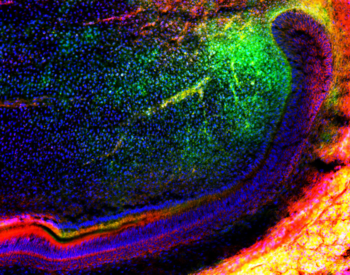
Stem cells hold the key to wound healing, as they develop into specialized cell types throughout the body—including in teeth. Now an international team of researchers, including KGI Associate Professor of Biopharmaceutical Sciences Alexander Zambon, has found a mechanism that could offer a potential novel solution to tooth repair.
Published August 9, 2019 in Nature Communications, the study showed that a gene called Dlk1 enhances stem cell activation and tissue regeneration in tooth healing. The work was led by Dr. Bing Hu from the University of Plymouth’s Peninsula Dental School, with collaboration from researchers worldwide—including Zambon and Tim Lee, a former Postbaccalaureate Premedical Certificate (PPC) student from KGI.
“With the help of Tim, we were able to provide Dr. Hu with a specialized genetically encoded sensor that facilitated the finding that Dlk1 caused dormant mesenchymal stem cells in teeth to awake from dormancy and divide,” Zambon said. “Dividing is a critical first step that enables the formation of the specialized cells (called transit amplifying cells) that are required to make each part of a tooth as it grows.”
The research team discovered a new population of mesenchymal stem cells (the stem cells that make up skeletal tissue such as muscle and bone) in a continuously growing mouse incisor model. They showed that these cells contribute to the formation of tooth dentin, the hard tissue that covers the main body of a tooth.
Importantly, the work showed that when these stem cells are activated, they then send signals back to the mother cells of the tissue to control the number of cells produced, through a molecular gene called Dlk1. This paper is the first to show that Dlk1 is vital for this process to work.
In the same report, the researchers also proved that Dlk1 can enhance stem cell activation and tissue regeneration in a tooth wound healing model. This mechanism could provide a novel solution for tooth reparation, dealing with problems such as tooth decay and crumbling (known as caries) and trauma treatment.
Further studies need to take place to validate the findings for clinical applications, in order to ascertain the appropriate treatment duration and dose, but these early steps in an animal model are exciting.
The full study is entitled Transit Amplifying Cells Coordinate Mouse Incisor Mesenchymal Stem Cell Activation and available to view now in Nature Communications.
The work was supported by the National Natural Science Foundation of China; National Institutes of Health; the Deutsche Forschungsgemeinschaft; the European Union Marie Skłodowska-Curie Actions; the European Regional Development Fund; and the Biotechnology and Biological Sciences Research Council.
Full list of institutions involved in the study:
- China: Peking University, Capital Medical University, Shenyang Stomatological Hospital and Second Hospital of Shandong University
- Denmark: University of Southern Denmark, University of Copenhagen
- Germany: Max Plank Institute for Molecular Biomedicine and Technische Universität Dresden
- Saudi Arabia: King Faisal University
- Singapore: National University of Singapore
- Switzerland: University of Geneva
- UK: University of Plymouth
- USA: Keck Graduate Institute and The University of Texas Health Science Center
